So, What Exactly is Electrospinning?
Basic Concepts

There are patents on electrospinning dating back to the 1930s, but not until recent years has it been applied a number of biomedical fields, including tissue engineering and drug delivery. The image to the left should give you a good idea of what a typical electrospinning set-up would look like. Electrospinning involves the use of a syringe needle (a bigger version of what you would see at the doctor's office) connected to a high-voltage (5 to 50 kilovolts) direct current power supply, a syringe pump to push the polymer out and a grounded collector plate (like foil). A polymer solution is loaded into the syringe, and this liquid is pushed from the needle tip at a constant rate by a syringe pump.
For a demonstration of electrospinning in action, check out this site !
A Bit of Physics...
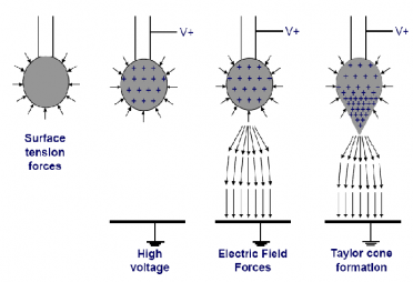
It seems relatively hard to believe that a solution in a syringe could be deposited onto a foil collector in such a uniform pattern, doesn't it? A lot of physics goes into understanding how this polymer solution changes forms from a relatively viscous (thick) liquid, to a mesh with microscopic holes. In the diagram to the right, you can see how the polymer solution forms a Taylor cone after leaving the tip of the syringe needle. The Taylor cone forms from the buildup of positive charge (+'s) at the tip of the solution droplet. Once the electrical field reaches a maximum value such that the forces that keep the droplet in its shape are overcome, a solution is ejected from the Taylor cone tip. As this jet travels toward the foil, the positive charges lead to the whirling of the jet as seen in the diagram above. This is then deposited on the foil collector plate as a mesh!
Understanding the Principal Force in Electrospinning

When the positive charges build up in the tip of the polymer droplet, forming the Taylor cone above, the electrical field eventually reaches a maximum value that allows the polymer to break free from its shape. The force the electrical field overcomes is called surface tension. Surface tension is the force that holds a group of molecules together, such as the water molecules that allow this bug to "walk" on their surface.
What Does an Electrospun Mesh Look Like?

An electrospun mesh, as shown here as a scanning electron micrograph (SEM), has the capability to be used in a number of biomedical applications because the pore size of a mesh can be changed by altering the components of the electrospinning set up:
- voltage
- syringe pump speed
- polymer type
- distance from the needle to the collector plate
Image below is a human hair in the foreground, with electrospun fibers in the back to give an idea about the scale. Image from:
http://vienna.bioengr.uic.edu/RET/Reports/Final%20Reports/ZufanRETFinalReport.pdf

